Cutaneous hemangiomas are benign growths that originate from endothelial cells (cells found in blood vessels) located in the skin and/or subcutaneous tissues. Hemangiosarcoma is a type of cancer that arises from blood vessels.

Hemangioma in Dogs What Is It and What to Do About It
A cutaneous hemangioma is a benign tumor found on the skin of dogs.

Photos of hemangiomas in dogs. They can grow on the surface or deeper into the center canal of a bone. Unfortunately, it’s not entirely known what causes this cancer in dogs. They are more common in dogs but do occur in cats, as well.
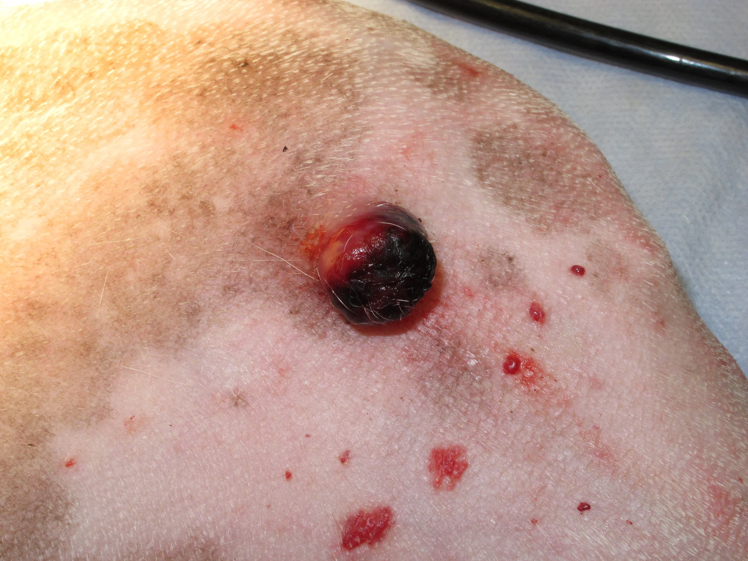
A benign tumor formed by a collection of excess blood vessels. Hemangiomas are single to multiple, circular, often compressible, red to black lumps and can look like a “blood blister.” Scientists are not 100 percent sure of what causes these skin growths to form.
If the tumor is detected early, and before it spreads to other tissue types within the body, surgical removal could result in a complete cure. For children, there may be a link between proteins developed while they are in the womb and hemangiomas. 6 in the dog, hemangiomas are typically benign, solitary, deep dermal tumors, whereas hemangiosarcomas often present as a disseminated malignancy involving the spleen,.
These tumors may be caused by certain chemicals, the sun, or they may be idiopathic (unknown cause) in origin. This form of cancer also seems to be more common in medium and large sized dog breeds, particularly: In addition, subcutaneous hemangiomas are typically elevated, partially hairless, and blue in color.
Hemangiomas in dogs are generally benign soft tissues and skin tumors, while sarcomas are malignant tumors developing in the soft tissues. Hemangiomas are small masses of blood vessels and are red to black in color. A hemangioma is a mass comprised of tangled blood vessels and connective tissue.
And airedale, scottish, and kerry blue terriers) are considered to be at risk. Carloni et al studied 61 dogs with visceral or muscular hsa diagnoses. Most hemangiomas that occur at birth disappear after a few months or years.
The color can vary from red to black, and the lesion can ulcerate. Hemangiomas are more noticeable in people with fair complexions. They are a bright red color.
Some vets may also recommend a whole body ct (computed tomographic) scan. Most cases of hsa have already spread at diagnosis. Risk factors for hemangiosarcoma may include:
Dermal hsa has the best prognosis. Hemangiomas are lesions of the vascular system that are formed by the cells that are responsible for forming blood vessels, or endothelial cells. Capillary hemangiomas are the most common type of hemangiomas.
Hemangiomas are capable of growing and becoming more prone to laceration, bruising, and infection. The hemangioma pictured here is of the simplest sort. These are rare tumors in cats, but common in dogs.
About one third of hemangiomas are present at birth. These images will show whether an hsa tumor has spread elsewhere in the body. Diet is an important consideration, although difficult to prove, a diet abundant in carbohydrates appears to encourage.
A hemangioma may be visible through the skin as a birthmark, known colloquially as a 'strawberry mark.'. Many breeds (including gordon setters; Unfortunately, hemangiosarcoma symptoms often don't become obvious until the cancer has already spread.
Congenital hemangiomas are present at birth. Animals that are overweight seem more disposed to growth growths. Any age or breed of dog can be afflicted, but hemangiosarcoma in dogs is most commonly seen in the breeds mentioned above, and in those over the age of six years.
Hemangiomas are benign tumors of adult dogs. Hemangiomas are formed by endothelial cells (the cells that form blood vessels). These benign tumors or growths vary in color from black to red.
Most hemangiomas are on the face and neck. Although uncommon, hemangiomas can develop in internal organs, most often the liver and intestines. A hemangioma is an abnormal buildup of blood vessels in the skin or internal organs.
Hemangiosarcomas can develop on the skin, under the skin in the subcutaneous tissue, and on internal organs such as the spleen and heart. The rest appear in the first several months of life. Male dogs also seem to have a higher chance of occurrence than female dogs.
The cause of hemangiomas in adults can be even more of a mystery. Spontaneous tumors of blood vessel endothelial cells have been described commonly in the dog, less frequently in the cat and horse, and sporadically in most other domestic species. Dermal, or cutaneous hemangiosarcoma, is the form of this canine cancer found in the skin.
There is some argument about whether these are even a cancer, although they are usually classified that way. Hemangiomas are a benign lump formed by blood vessel tissues that occur in the skin. However, hemangiosarcoma has been found in puppies less than year old.
Hemangiomas occur in a variety of sizes, locations, and degrees of combination of capillaries, larger vessels, and venous lakes. It presents as a red or black growth on the skin. Hemangiosarcoma, or cancer of the blood vessels, is a type of tumor occurring more often in canines than any other animal.
Especially if your dog is lame. That makes sense, because hemangiomas are vascular lesions, formed by endothelial cells, which are the cells that form blood vessels. The cause of hemangiomas is idiopathic (unknown).
It originates either in the dermis or the subcutaneous layer of the dog’s skin. These tumors are most common on the legs and trunk. Hemangiosarcoma is a cancer that starts in the endothelial cells which are the cells that line the blood vessel walls.
A hemangioma can grow, making it prone to bruising, laceration, and infection.
Skin Tumors HSA — DR. JULIUS LIPTAK

Dante the Dog's Cutaneous Hemangioma Laser Surgery Story
What Dog Skin Cancer Looks Like Signs, Pics

[PDF] Cutaneous in a Dog Semantic Scholar

My dog has a few black dots on his belly and one is a red